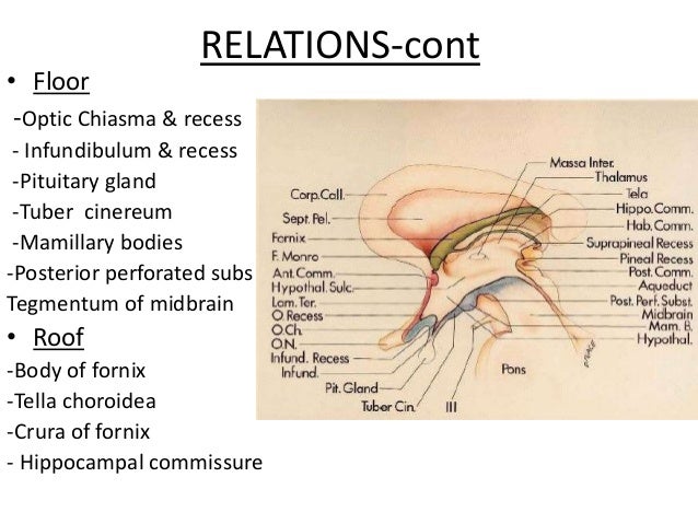
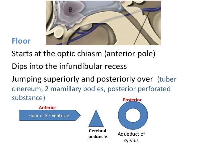
Floor Of Third Ventricle Perferated

Third Ventricle Location Boundaries Recesses And Choroid Plexus Anatomy Qa

3rd Ventricle N Pineal Gland
Floor Of Third Ventricle Mnemonics
Floor Of 3rd Ventricle Form By Mnemonics
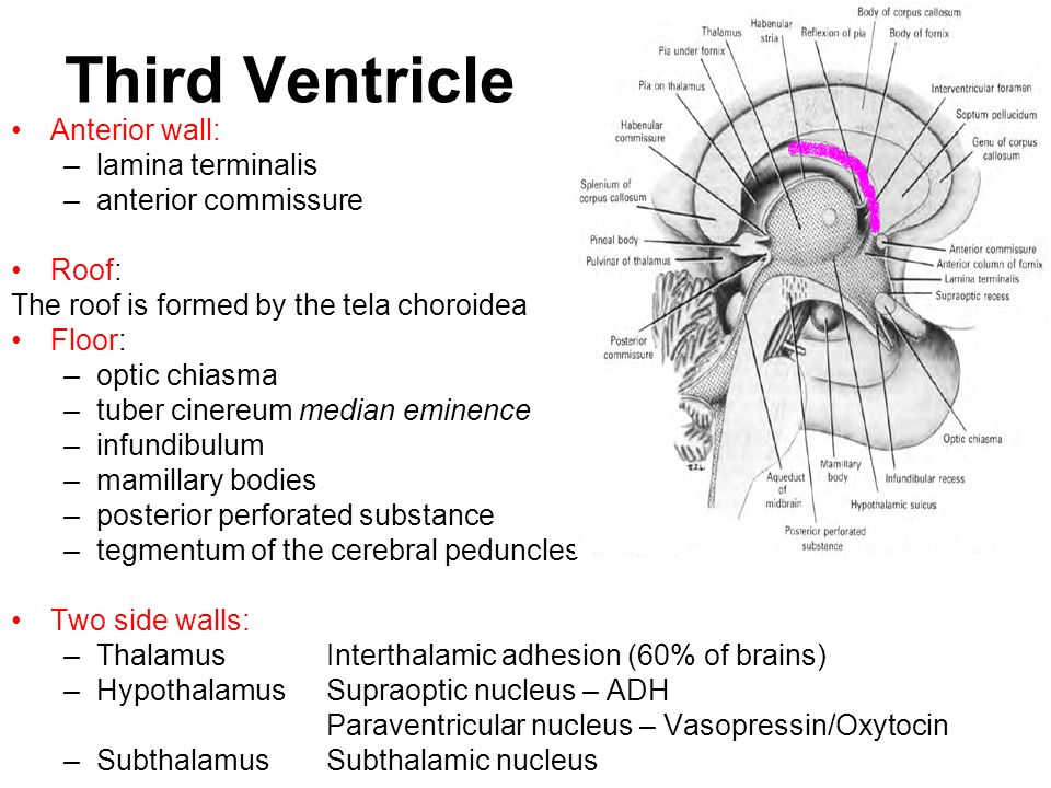
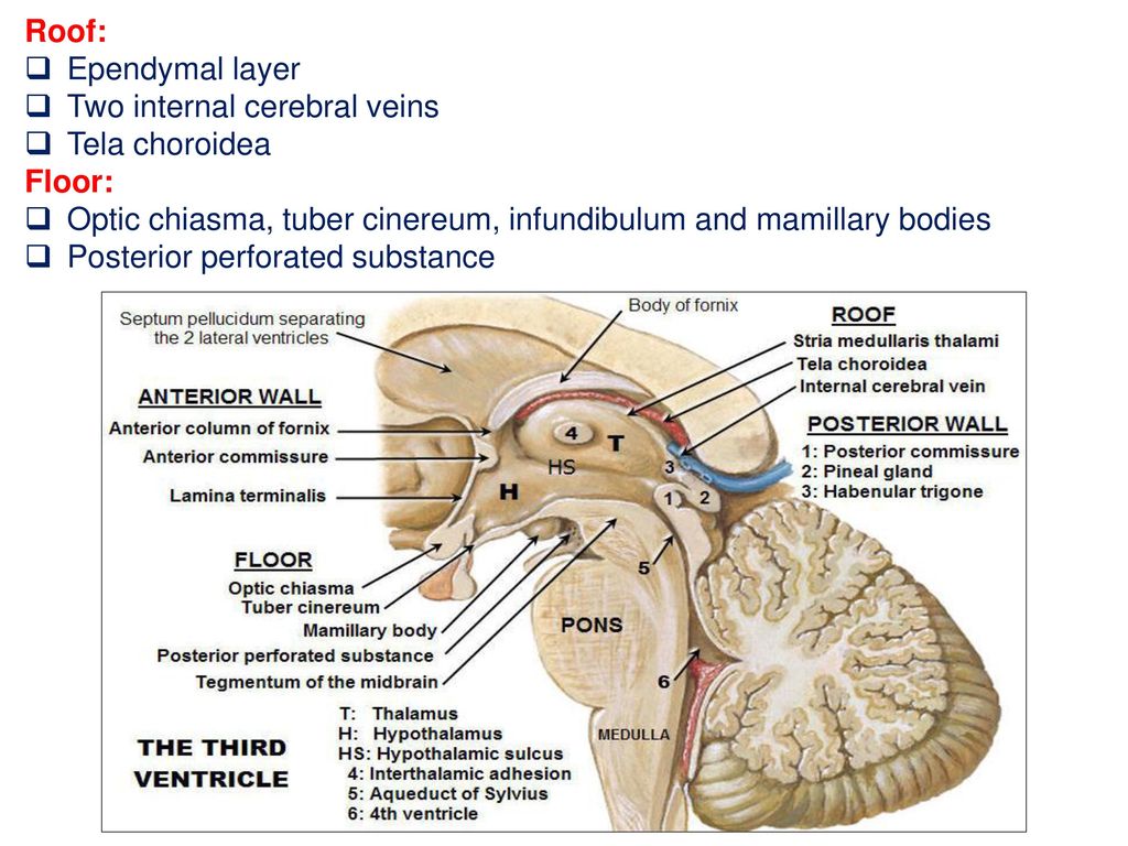
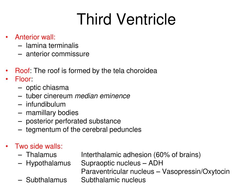
Third Ventricle
Brain Anatomy
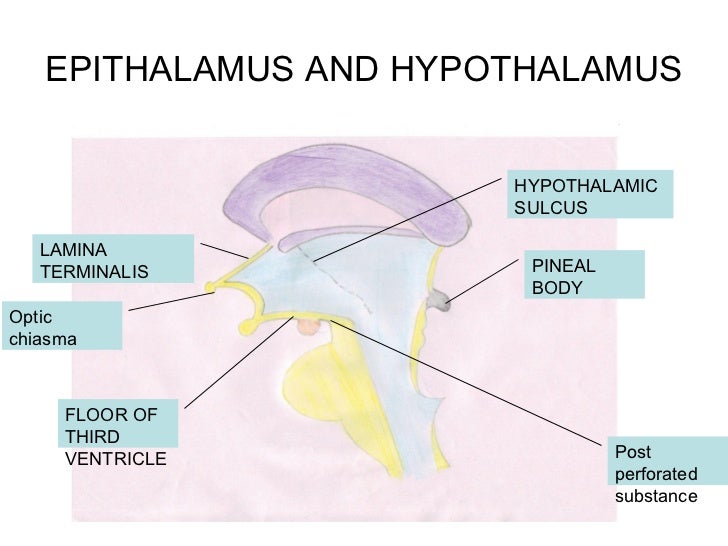
Anterior wall is formed from above downwards by.
Floor of third ventricle perferated.

Pathology Of Hypothalamus Pituitary I Dr Jones Flashcards Quizlet
Third Ventricle
Floor And Roof Of The Third Ventricle Neuroanatomy The Neurosurgical Atlas By Aaron Cohen Gadol M D
Meninges Csf Ventricular System Objectives Describe The Arrangement Of The Meninges And Their Relationship To Brain And Spinal Cord Explain The Occurrence Ppt Download

Approaches To Third Ventricular Tumors Sciencedirect
Meninges Csf And Ventricular System Ppt Download
Third Ventricle

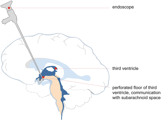
Endoscopic View Of Lm Following Traversing The Floor Of The Third Download Scientific Diagram

Pdf Histological Analysis Of The Third Ventricle Floor In Hydrocephalic And Nonhydrocephalic Brains Application To Neuroendocrine Complications Following Third Ventriculostomy Procedures Laboratory Investigation

Embryology Of Ventricles Closure Of Neural Tube By 28 Days Of Gestation Certain Portions Of Central Lumen Expand To Form Basic Pattern Of Ventricular Ppt Download

Jaypeedigital Ebook Reader

An Overview Of The Third Ventricle Floor Ventral View Using A Ruler Download Scientific Diagram

81 Third Ventricle الدكتور أحمد كمال المقطع الأخير Youtube

Third Ventricle Anatomy Kenhub
Pdf Third Ventricle
Meninges Csf Ventricular System Ppt Download
Neurosurgical Considerations In Macrocephaly Springerlink

Jules Musings 4 Years
Https Encrypted Tbn0 Gstatic Com Images Q Tbn 3aand9gcrh A1aidtnjkifzkxi6ilqzeeni1wbdie9qtmj4t7j Te1h7bj Usqp Cau
Http Website60s Com Upload Files 5 Pseudo Spontaneous Third Ventriculostomy Pdf

Comparing The Efficiency Of Two Treatment Methods Of Hydrocephalus Shunt Implantation And Endoscopic Third Ventriculostomy Basic And Clinical Neuroscience

Pdf Endoscopic Third Ventriculostomy
T7p1hm0k8x1dsm
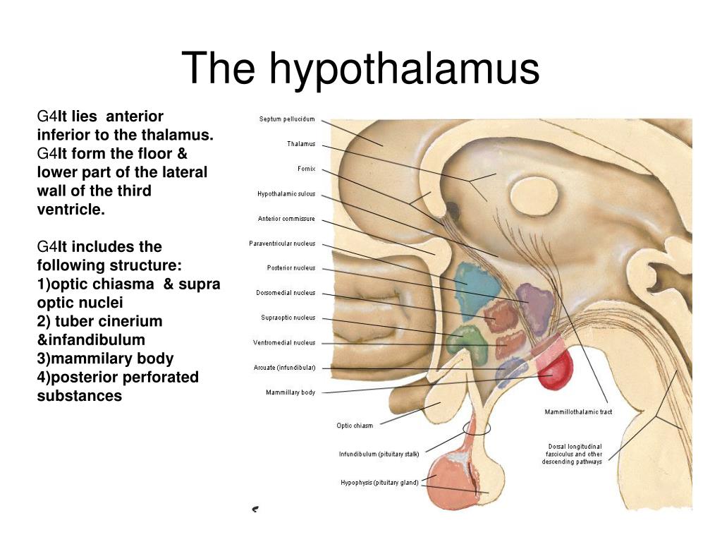
Ppt Diencephalon Thalamus Hypothalamus Powerpoint Presentation Id 3830362
Source : pinterest.com
